Authors: Xiang Zhang, Bryan Ranger, Jon Fincke, Hugh Herr, Brian Anthony
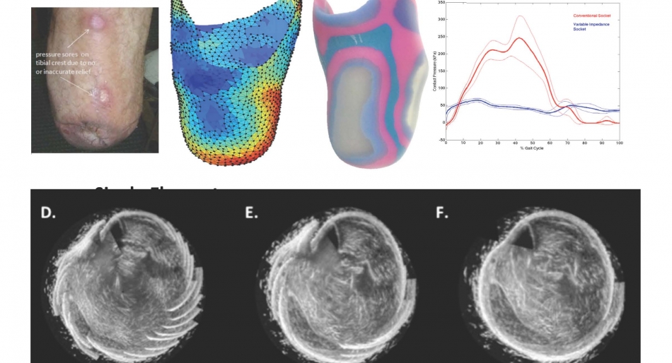
The quality of a prosthetic fitting is determined by the biomechanical interaction between the prosthetic socket and the residual limb. An optimized fitting can improve muscle usage, relieve pressure points, and improve overall mobility. However, current methods of prosthetic socket design remain subjective and ineffective at creating a comfortable and functional fitting. Though magnetic resonance imaging (MRI) and X-ray computed tomography (CT) have been explored to improve biomechanical modeling for fittings, a low-cost, streamlined, and quantitative process has not been proven. Ultrasound, already used extensively for imaging and diagnostics, has shown promise in capturing the internal and external limb geometries required for improved biomechanical modeling. Recent increases in computational power have enabled processing of transmission and backscatter data on clinically relevant time scales. As a result, ultrasound tomography systems are now being developed for clinical use. Here, we present the development of a single element scanning system with corresponding reconstruction algorithms and an array scanning system with corresponding image stitching techniques to capture the internal and external geometries of proximal limbs.